Xenopus laevis: Difference between revisions
imported>Daniel Mietchen (+image) |
mNo edit summary |
||
| (2 intermediate revisions by 2 users not shown) | |||
| Line 6: | Line 6: | ||
| trend = up | | trend = up | ||
--> | --> | ||
| image = Xenopus_laevis. | | image = Xenopus_laevis.png | ||
| image_width = 240px | | image_width = 240px | ||
| regnum = [[Animal]]ia | | regnum = [[Animal]]ia | ||
| Line 32: | Line 32: | ||
They also swim very fast, and can eat smaller fish, such as minnows and guppies. When they eat, the food is not actually held down very well, and occasionally, the fish, or bug, can actually escape before being forced back into the mouth by the frog's front hands. | They also swim very fast, and can eat smaller fish, such as minnows and guppies. When they eat, the food is not actually held down very well, and occasionally, the fish, or bug, can actually escape before being forced back into the mouth by the frog's front hands. | ||
{{Image|Xenopus laevis tadpole nervous system.png|right|350px|Schematic overview of the [[nervous system]] of a hatchling X. laevis: (a) Photograph of tadpole at [[developmental stage|stage]] 37/38. (b) The main parts of the [[central nervous system]] with arrowhead at [[hindbrain]]/[[spinal cord]] border. (c) Transverse section of the spinal cord with the left side [[immunohistochemistry|stained]] to show [[glycine]] immunoreactive [[cell body|cell bodies]] (arrows) and | {{Image|Xenopus laevis tadpole nervous system.png|right|350px|Schematic overview of the [[nervous system]] of a hatchling X. laevis: (a) Photograph of tadpole at [[developmental stage|stage]] 37/38. (b) The main parts of the [[central nervous system]] with arrowhead at [[hindbrain]]/[[spinal cord]] border. (c) Transverse section of the spinal cord with the left side [[immunohistochemistry|stained]] to show [[glycine]] immunoreactive [[cell body|cell bodies]] (arrows) and axons (in the marginal zone). Diagrammatic right side shows the main regions: [[neural canal]] (c) bounded by [[ventral floor plate]] (f) and [[ependym]]al cell layer (e); [[lateral marginal zone]] of axons (mauve), layer of differentiated neuron cell bodies arranged in longitudinal columns (coloured circles) lying inside the marginal zone except in dorso-lateral (dl) and dorsal positions. (d) Diagrammatic view of the spinal cord seen from the left side, showing characteristic position and features of seven different neuron types. Each has a [[soma]] (solid ellipse), [[dendrite]]s (thick lines) and axon(s) (thin lines). [[Commissural]] axons [[neuronal projection|projecting]] on the opposite right side are dashed.}} | ||
== Use in genetic research == | == Use in genetic research == | ||
| Line 53: | Line 53: | ||
==References== | ==References== | ||
{{reflist}} | {{reflist}} | ||
[[Category:Suggestion Bot Tag]] | |||
Latest revision as of 17:00, 9 November 2024
- The content on this page originated on Wikipedia and is yet to be significantly improved. Contributors are invited to replace and add material to make this an original article.
| African clawed frog | ||||||||||||||
|---|---|---|---|---|---|---|---|---|---|---|---|---|---|---|
 | ||||||||||||||
| Scientific classification | ||||||||||||||
| ||||||||||||||
| Binomial name | ||||||||||||||
| Xenopus laevis Daudin, 1802 |
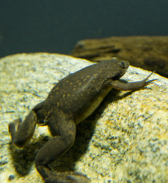
The African clawed frog (Xenopus laevis, also known as platanna) is a species of South African aquatic frog of the genus Xenopus. It can grow up to 12 cm long with a flattened head and body, but no external ear or tongue. Its name derives from the three short claws on each of its hind feet, which it probably uses to stir up mud to hide it from predators.
The species is found throughout much of Africa, and in isolated, introduced populations in North America, South America, and Europe.[1]
Description
These frogs are plentiful in ponds and rivers within the southeastern portion of Sub-Saharan Africa. They are aquatic and often a yellowish, grey color. Albino varieties are sold as pets. They reproduce by eggs (see frog reproduction).
These frogs tend to live 5 to 15 years but some have been recorded to live to be nearly 30 years. They shed every season, and eat their own shed skin.
Although lacking a vocal sac, the males make a mating call of alternating long and short trills, by contracting the intrinsic laryngeal muscles. Most unusually, females also answer vocally, signaling either acceptance (a rapping sound) or rejection (slow ticking) of the male.[2]
They also swim very fast, and can eat smaller fish, such as minnows and guppies. When they eat, the food is not actually held down very well, and occasionally, the fish, or bug, can actually escape before being forced back into the mouth by the frog's front hands.

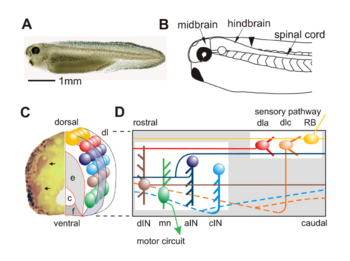
Schematic overview of the nervous system of a hatchling X. laevis: (a) Photograph of tadpole at stage 37/38. (b) The main parts of the central nervous system with arrowhead at hindbrain/spinal cord border. (c) Transverse section of the spinal cord with the left side stained to show glycine immunoreactive cell bodies (arrows) and axons (in the marginal zone). Diagrammatic right side shows the main regions: neural canal (c) bounded by ventral floor plate (f) and ependymal cell layer (e); lateral marginal zone of axons (mauve), layer of differentiated neuron cell bodies arranged in longitudinal columns (coloured circles) lying inside the marginal zone except in dorso-lateral (dl) and dorsal positions. (d) Diagrammatic view of the spinal cord seen from the left side, showing characteristic position and features of seven different neuron types. Each has a soma (solid ellipse), dendrites (thick lines) and axon(s) (thin lines). Commissural axons projecting on the opposite right side are dashed.
Use in genetic research
Although X. laevis does not have the short generation time and genetic simplicity generally desired in genetic model organisms, it is an important model organism in developmental biology. X. laevis takes 1 to 2 years to reach sexual maturity and, like most of its genus, it is tetraploid. However, it does have a large and easily manipulable embryo. The ease of manipulation in amphibian embryos has given them an important place in historical and modern developmental biology. A related species, Xenopus tropicalis, is now being promoted as a more viable model for genetics. Roger Wolcott Sperry used X. laevis for his famous experiments describing the development of the visual system. These experiments led to the formulation of the Chemoaffinity hypothesis.
Xenopus oocytes provide an important expression system for molecular biology. By injecting DNA or mRNA into the oocyte or developing embryo, scientists can study the protein products in a controlled system. This allows rapid functional expression of manipulated DNAs (or mRNA). This is particularly useful in electrophysiology, where the ease of recording from the oocyte makes expression of membrane channels attractive. One challenge of oocyte work is eliminating native proteins that might confound results, such as membrane channels native to the oocyte. Translation of proteins can be blocked or splicing of pre-mRNA can be modified by injection of Morpholino antisense oligos into the oocyte (for distribution throughout the embryo) or early embryo (for distribution only into daughter cells of the injected cell).[3]
X. laevis is also notable for its use as the first well-documented method of pregnancy testing when it was discovered that the urine from pregnant women induced X. laevis oocyte production. Human chorionic gonadotropin (HCG) is a hormone found in substantial quantities in the urine of pregnant women. Today, commercially available HCG is injected into Xenopus males and females to induce mating behavior and breed these frogs in captivity at any time of the year.
As a pest
When African clawed frogs are imported into non-native countries, they have the capacity to wreck entire ecosystems by eating native wildlife such as fish and turtles that have no natural defense against these creatures.
In 2007, these frogs invaded a pond in San Francisco, where much debate exists on how to terminate these creatures and keep them from spreading.[4][5] It is unknown if these frogs entered the San Francisco ecosystem through intentional release or escape into the wild.
Because these frogs are immune to the fungi Batrachochytrium dendrobatidis (a chytridomycota) and B. dendrobatidis has been traced back to the habitat of Xenopus laevis in Africa, many scholars believe it is the source of the worldwide frog population crash. Due to its extensive use in obstetrics and research, it appears Xenopus laevis has carried B. dendrobatidis with it out of Africa to all over the world, causing chytridomycosis and eventually death in native frogs naïve to the fungi.
As a pet
These frogs are increasing in popularity on the home aquarium market due to the fact the they are relatively easy to keep as pets. They are usually bred in community aquariums with other fish or aquatic frogs larger than itself. They are fed bloodworms, shrimp, and brine shrimp either live or dried.
References
- ↑ Tinsley et al (2004). Xenopus laevis. 2006 IUCN Red List of Threatened Species. IUCN 2006. Retrieved on 12 May 2006. Database entry includes a range map and justification for why this species is of least concern
- ↑ ADW: Xenopus Laevis: Information
- ↑ Comparison of morpholino based translational inhib...[Genesis. 2001] - PubMed Result
- ↑ Killer Meat-Eating Frogs Terrorize San Francisco. FoxNews.
- ↑ The Killer Frogs of Lily Pond:San Francisco poised to checkmate amphibious African predators of Golden Gate Park. San Francisco Chronicle.